


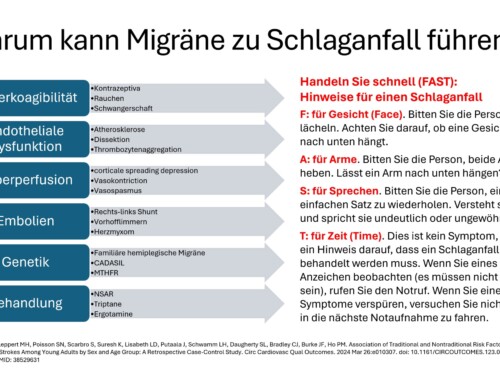
There are only a few effective treatment options for particularly severely affected patients with chronic migraines and other severe chronic pain. Standard therapy procedures are usually without lasting effectiveness. Recently, peripheral nerve stimulation (PNS) has come into focus as a treatment option. Peripheral nerve stimulation is a specific application of neuromodulation. This has been used to relieve chronic pain for several decades. Successful use is possible for headaches, back pain, neck pain, arm and leg pain. Due to the increasing progress of microelectronics, it is possible to implant a pacemaker-like device under the skin and thus enable continuous neuromodulation. The device is about the size of a matchbox.
To treat chronic migraines, a special rechargeable system can be implanted, meaning there is no need to change the battery. The stimulator sends electrical signals to the occipital nerve (ON), located just under the skin of the neck. Due to this special location, the treatment option is also called occipital nerve stimulation (ONS). The mode of action of occipital nerve stimulation is explained by changes in electrical regulation in the brainstem. The pattern of pain signals is modulated and masked by continuous stimulation. The constant hypersensitivity in the nervous system is balanced and reduced. The function of the neurostimulator system and the peripheral nerve stimulation can be compared to that of the pacemaker. It is assumed that neuromodulation activates and stabilizes the body's own defenses against pain, thus naturally reducing sensitivity to pain signals. The individual steps of implanting the system are described below.
For the decision to undergo neuromodulation, the patient must be cared for in a specialized migraine and headache center. The specialized indication and further professional care must be guaranteed. In our center, in collaboration with the neurosurgery clinics of the University Hospital Schleswig-Holstein, the process described below has proven successful.
The indication and information about treatment options are provided in the Migraine and Headache Center at the Kiel Pain Clinic. We will then organize an outpatient visit to the neurosurgery clinic to plan the operation and anesthesia. Due to our nationwide headache treatment network, care may also be provided at another cooperating and certified neurosurgical center.
The stimulator is implanted on the planned date, usually as follows:
Day 1: Admission to neurosurgery the evening before
Day 2: Implantation in the morning
Day 3: Initial setting of the stimulation parameters in the afternoon
Day 4: Discharge in the morning
As a rule, the stimulator is then fine-tuned in the Kiel Pain Clinic:
Day 1: Admission after discharge from neurosurgery
Day 2: Checking the stimulation parameters, resetting the medication
Day 3: Discharge
The implantation is usually carried out according to the following description:
Induction of anesthesia, the patient is intubated and positioned. During the operation, the patient lies on his stomach with his head slightly bent downwards or forwards. The exact position of the electrodes is then recorded from the outside using a thin rod under X-ray control and marked on the outside of the skin. A marking is made on both sides of the ear area from the outside so that you know exactly where the electrode should be located. The hair on the neck area is shaved and possibly the back is shaved for hairy men. The skin areas are then marked to locate the stimulator.
After disinfection and covering, the skin is opened in the area of the occipital nerve through a longitudinal incision and coagulation of the small blood vessels as well as the opening of the small pockets on the left and right for the cables. The 8-pole electrodes, which are held on a metal rod, are then measured from the outside. This stick is now pushed on one side directly under the skin towards the ear, according to the mark previously made. If you pull it back, a small plastic tube remains under the skin. The first electrode is now pushed through this. If it sits perfectly (the location had already been marked from the outside), the plastic tube is pulled back and the electrode remains in place. The second electrode is placed on the other side in the same way. The cables are then sewn to the fascia so that they cannot slip.
In the next step, a longer, thin metal tube is used to pave the way from the occipital nerve to the end of the back, through which the flexible cables are then passed. If you pull the metal tube back again, the small plastic tube remains through which the cables are pushed. If they fit perfectly, the plastic tube is pulled back and only the thin cables remain under the skin.
We prefer to use the stimulator on the lower back below the belt line. A flexible extension (cable extension) is installed above the belt line so that the cables have enough play and cannot tear or break. The cables are now connected to each other, the cable parts are sewn to the fascia so that they cannot slip, and the stimulator is inserted. Before the skin pocket is sutured, the technician checks all individual values very carefully. If all values are perfect, the wounds are closed subcutaneously with absorbable material. The opening on the neck is also closed on the outside with absorbable material; the two openings on the back are stapled if necessary.
An X-ray check is then carried out to check the position of the electrodes and the stimulator.
The patient wakes up and is taken to the room. The technician comes in the afternoon or the next day, explains the procedure in detail to the patient and the first adjustment is made.
Further care and fine-tuning of the stimulator are then carried out by the Migraine and Headache Center at the Kiel Pain Clinic. After transfer to our clinic, the functionality is checked and the stimulation pattern is individually adjusted and adjusted. The setting is done externally via a small transmitter that transmits the values to the stimulator and can control them. Depending on the situation, an optimized pattern for neuromodulation can be set. The patient also receives a transmitter to control the device independently. For this purpose, various settings are pre-programmed, which the patient can then access individually. Finally, the medication is checked and, if necessary, adjusted again. The patient can then be discharged home and can then present themselves for further follow-up checks.


[…] can be reduced. The individual steps of implanting the system and further details can be found here. This article was posted under General, Cluster Headache, Cluster Headache Competence Center, […]